Unveiling the Human Abdomen: A Comprehensive Guide to its Regions and Structures
Related Articles: Unveiling the Human Abdomen: A Comprehensive Guide to its Regions and Structures
Introduction
In this auspicious occasion, we are delighted to delve into the intriguing topic related to Unveiling the Human Abdomen: A Comprehensive Guide to its Regions and Structures. Let’s weave interesting information and offer fresh perspectives to the readers.
Table of Content
- 1 Related Articles: Unveiling the Human Abdomen: A Comprehensive Guide to its Regions and Structures
- 2 Introduction
- 3 Unveiling the Human Abdomen: A Comprehensive Guide to its Regions and Structures
- 3.1 Mapping the Abdominal Landscape: A Regional Approach
- 3.2 Anatomical Structures within the Abdominal Map:
- 3.3 Significance of the Abdominal Map:
- 3.4 Frequently Asked Questions (FAQs) about the Abdominal Map:
- 3.5 Tips for Understanding the Abdominal Map:
- 3.6 Conclusion:
- 4 Closure
Unveiling the Human Abdomen: A Comprehensive Guide to its Regions and Structures
The human abdomen, the central region of the body between the thorax (chest) and the pelvis, serves as a vital hub for numerous physiological functions. Its intricate network of organs, muscles, and vessels plays a critical role in digestion, nutrient absorption, waste elimination, and the overall well-being of the individual. Understanding the anatomical map of the abdomen is crucial for healthcare professionals, students, and anyone seeking a deeper knowledge of the human body.
Mapping the Abdominal Landscape: A Regional Approach
To navigate the complex landscape of the abdomen, it is essential to divide it into distinct regions. This regional approach provides a structured framework for understanding the location and function of various organs and structures.
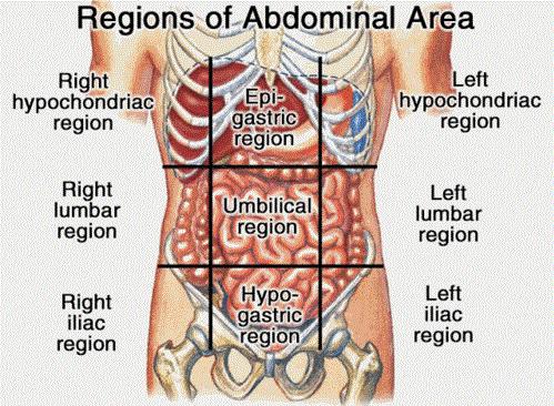
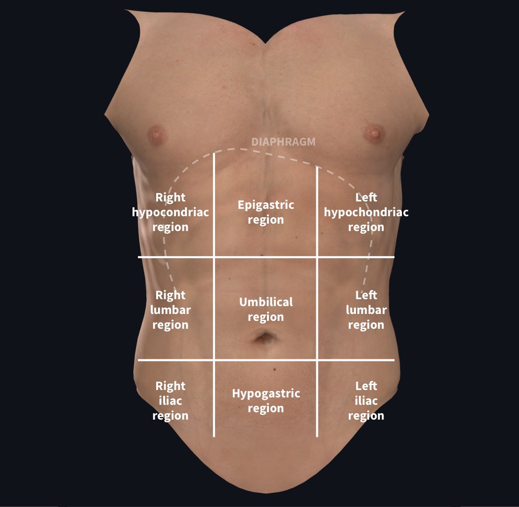
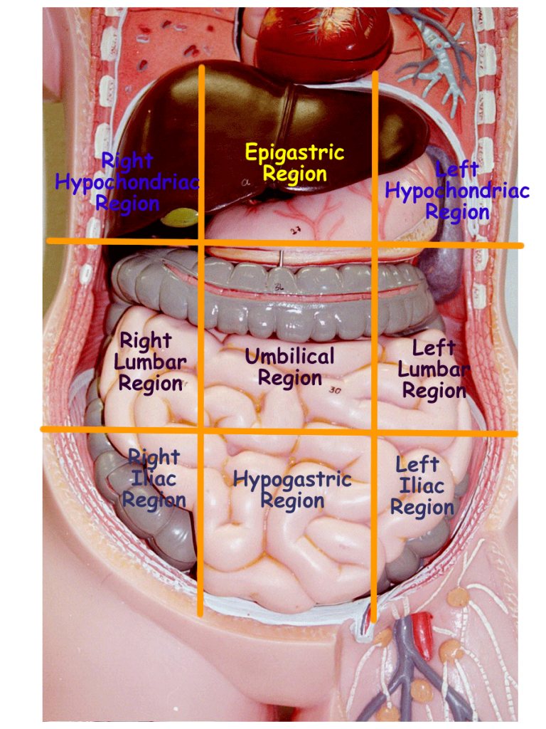
1. Nine Regions of the Abdomen:
The nine-region system is widely used in clinical settings and anatomical studies. It divides the abdomen into three horizontal and three vertical lines:
-
Horizontal Lines:
- Subcostal line: Passes horizontally across the lower border of the tenth rib.
- Transpyloric line: Passes horizontally through the pylorus of the stomach, typically at the level of the first lumbar vertebra.
- Intertubercular line: Passes horizontally across the iliac tubercles, marking the upper border of the pelvis.
-
Vertical Lines:
- Midclavicular lines: Pass vertically down from the midpoint of each clavicle.
These lines intersect to define nine distinct regions:
-
Upper Row:
- Right hypochondriac region: Houses the liver, gallbladder, and part of the right kidney.
- Epigastric region: Contains the stomach, duodenum, pancreas, and part of the liver.
- Left hypochondriac region: Encompasses the spleen, left kidney, and part of the stomach.
-
Middle Row:
- Right lumbar region: Includes the ascending colon, part of the right kidney, and the right ureter.
- Umbilical region: Located around the umbilicus, contains the small intestine, part of the transverse colon, and the abdominal aorta.
- Left lumbar region: Contains the descending colon, part of the left kidney, and the left ureter.
-
Lower Row:
- Right iliac region: Houses the cecum, appendix, and part of the right ovary or spermatic cord.
- Hypogastric region: Encompasses the urinary bladder, uterus (in females), and the sigmoid colon.
- Left iliac region: Contains the sigmoid colon, part of the left ovary or spermatic cord.
2. Four Quadrants of the Abdomen:
A simpler approach divides the abdomen into four quadrants using two perpendicular lines:
- Midline: Runs vertically down the center of the abdomen.
- Transumbilical line: Passes horizontally through the umbilicus.
These lines create four quadrants:
- Right upper quadrant (RUQ): Contains the liver, gallbladder, part of the pancreas, and the right kidney.
- Left upper quadrant (LUQ): Houses the stomach, spleen, left kidney, and part of the pancreas.
- Right lower quadrant (RLQ): Includes the appendix, cecum, part of the ascending colon, and the right ovary or spermatic cord.
- Left lower quadrant (LLQ): Contains the descending colon, sigmoid colon, and the left ovary or spermatic cord.
Anatomical Structures within the Abdominal Map:
The abdominal map is not merely a collection of regions; it serves as a guide to understanding the complex interplay of organs and structures within the abdomen.
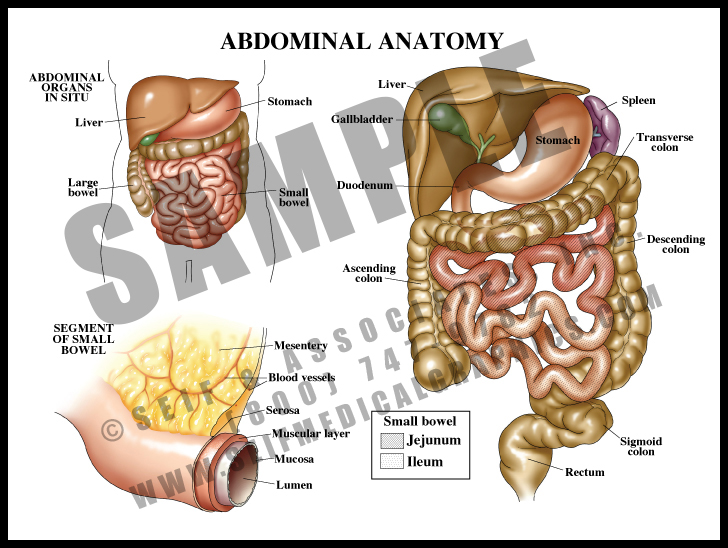
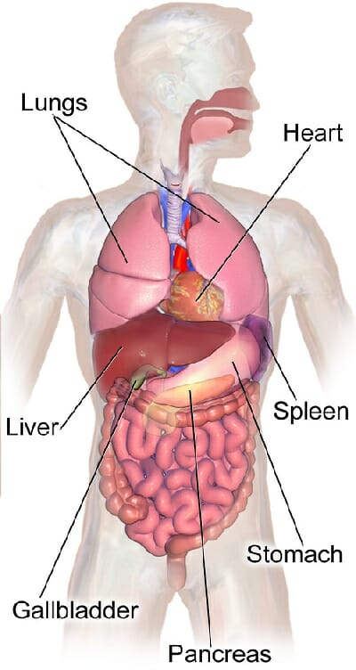
1. Digestive System:
The digestive system, responsible for breaking down food and absorbing nutrients, dominates the abdominal cavity. Key organs include:
- Stomach: A muscular sac that churns and mixes food with gastric juices.
- Small intestine: A long, coiled tube where most nutrient absorption occurs.
- Large intestine: A shorter, wider tube responsible for water absorption and waste elimination.
- Liver: The largest internal organ, responsible for detoxification, bile production, and nutrient processing.
- Gallbladder: Stores and concentrates bile, aiding in fat digestion.
- Pancreas: Secretes digestive enzymes and hormones that regulate blood sugar levels.
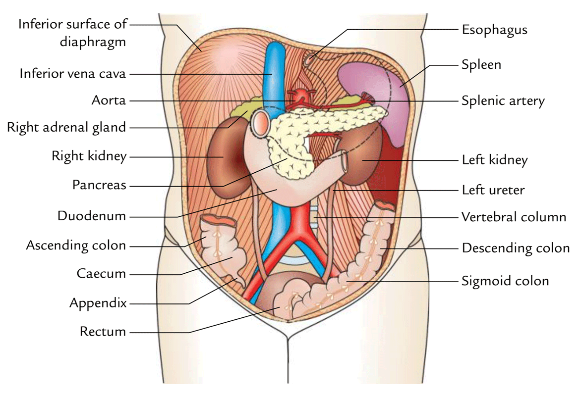
2. Urinary System:
The urinary system, responsible for filtering waste products from the blood and producing urine, also plays a significant role in maintaining fluid balance. Key organs include:
- Kidneys: Filter waste products from the blood and produce urine.
- Ureters: Carry urine from the kidneys to the bladder.
- Bladder: Stores urine until it is expelled.
3. Reproductive System:
The reproductive system, responsible for sexual reproduction, is housed within the pelvic cavity, which is considered a continuation of the abdominal cavity. Key organs include:
- Ovaries (females): Produce eggs and hormones.
- Uterus (females): Houses a developing fetus.
- Fallopian tubes (females): Transport eggs from the ovaries to the uterus.
- Testes (males): Produce sperm and hormones.
4. Other Structures:
The abdominal cavity also contains other vital structures:
- Spleen: Filters blood, stores blood cells, and plays a role in immunity.
- Abdominal aorta: The main artery supplying blood to the abdomen and lower extremities.
- Inferior vena cava: The main vein returning blood from the lower body to the heart.
- Peritoneum: A serous membrane lining the abdominal cavity and covering the abdominal organs.
Significance of the Abdominal Map:
Understanding the abdominal map is crucial for various reasons:
1. Medical Diagnosis:
- Physical examination: Physicians use the abdominal map to guide their physical examination, allowing them to palpate organs, listen to bowel sounds, and assess tenderness.
- Imaging studies: Imaging techniques like ultrasound, CT scans, and MRI rely on anatomical landmarks to visualize and diagnose abdominal conditions.
- Surgical procedures: Surgeons use the abdominal map to plan and execute surgical procedures with precision.
2. Patient Education:
- Understanding symptoms: Patients can better understand their symptoms when they know the location of the organs involved.
- Communicating with healthcare providers: Patients can communicate more effectively with their healthcare providers by using accurate anatomical terminology.
- Promoting self-care: Understanding the abdominal map can empower individuals to take proactive steps towards their health, such as maintaining a healthy diet and lifestyle.
3. Research and Development:
- Anatomical studies: The abdominal map serves as a foundation for anatomical research, enabling scientists to study the structure and function of abdominal organs.
- Drug development: Pharmaceutical companies use the abdominal map to understand the target organs for new drugs and therapies.
- Medical devices: Engineers design medical devices, such as catheters and stents, based on the anatomical map of the abdomen.
Frequently Asked Questions (FAQs) about the Abdominal Map:
1. What is the most common cause of abdominal pain?
Abdominal pain can have numerous causes, ranging from indigestion and gas to serious conditions like appendicitis or pancreatitis. It’s important to consult a healthcare professional for a proper diagnosis and treatment plan.
2. How can I prevent abdominal pain?
Maintaining a healthy diet, staying hydrated, managing stress, and engaging in regular physical activity can help prevent abdominal pain.
3. What are the symptoms of a ruptured appendix?
Symptoms of a ruptured appendix include severe abdominal pain, nausea, vomiting, fever, and loss of appetite. Seek immediate medical attention if you suspect a ruptured appendix.
4. What are the benefits of a healthy gut microbiome?
A healthy gut microbiome plays a crucial role in digestion, nutrient absorption, immunity, and overall health.
5. What is the difference between the nine regions and four quadrants of the abdomen?
The nine-region system provides a more detailed anatomical map, while the four-quadrant system offers a simpler, more practical approach for clinical purposes.
Tips for Understanding the Abdominal Map:
- Visual aids: Use anatomical diagrams, models, and online resources to visualize the abdominal map.
- Labeling exercises: Practice labeling the regions and organs of the abdomen on diagrams.
- Clinical correlation: Relate the anatomical map to clinical scenarios and patient symptoms.
- Hands-on learning: Engage in physical examination techniques to familiarize yourself with the location of abdominal organs.
- Continuous learning: Stay updated on the latest advancements in abdominal anatomy and related medical fields.
Conclusion:
The abdominal map is an essential tool for understanding the human body. It provides a framework for comprehending the complex anatomy and physiology of the abdomen, facilitating accurate diagnosis, effective treatment, and informed patient care. By mastering the abdominal map, individuals can gain a deeper appreciation for the intricate workings of their bodies and make informed decisions about their health and well-being.


Closure
Thus, we hope this article has provided valuable insights into Unveiling the Human Abdomen: A Comprehensive Guide to its Regions and Structures. We appreciate your attention to our article. See you in our next article!